
WHAT IS A MAST CELL?
Mast cells are found in all tissues of the body. This cell is part of our immunologic defense systems against invading organisms but they are in especially high numbers in the skin, respiratory tract, and gastrointestinal tract.
Inside the mast cell are tiny granules containing different chemicals that cause inflammation; mostly histamines (causing leakage of fluid resulting in swelling and itching) and heparin (a blood thinner), meant for use against invading parasites. Think of these as small bombs that can be released. They play a role in allergic responses, non-allergic skin disease, wound healing and tissue remodeling. They can also increase stomach acid production.
These unique cells control many bodily functions and are important immune, allergy and infection fighting or defensive cells. Mast cells are meant to participate in the war against parasites (as opposed to the war against bacterial or viral invaders). They are bound within tissues that interface with the external world such as the skin, respiratory or intestinal tract. They do not circulate through the body.When mast cells replicate in higher than normal numbers, a mast cell tumor can form. Pets with mast cell tumors can also have complications like stomach problems from the overproduction of stomach acid and excessive bleeding from the release of heparin.
Mast cell tumors occur in both dogs and cats. Although some are benign, most of these tumors are malignant. Mast cell tumors vary widely in their appearance, shape, and size.
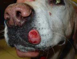
If your pet has a mast cell tumor on the skin, there will be a bump or lesion of some kind. Sometimes it's a raised pink bump on the surface of the skin. Sometimes it's a less-defined mass that feels like a lump under the skin.
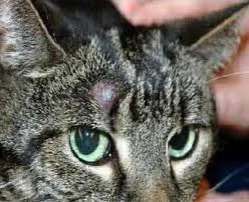
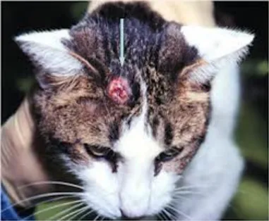
Mast cell tumors in cats are usually
solitary and are found on the head.
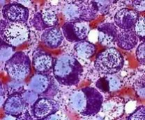
Mast Cells with dark granules
AND THE MAST CELL TUMOR?
The mast cell can form a tumor made of many mast cells either in the skin or in deeper tissues like connective tissue between muscles. When this happens, the cells of the tumor are unstable. This means they release their toxic granules with simple contact or even at random, creating allergic symptoms that do not correlate with exposure to any particular antigen. The release of histamines can result in severe swelling of the nearby tissues and intense itching.
Mast Cell Tumors in Dogs
Mast cell tumors are notoriously invasive and difficult to treat.
Between 7 and 21% of all canine skin tumors are mast cell tumors.
Mast cell tumors are especially common in dogs accounting for approximately one skin tumor in every five. The Boxer is at an especially high risk, as are related breeds: English Bulldog, Boston Terrier. Also at higher than average risk are the Shar pei, Labrador Retriever, Golden Retriever, Schnauzer, and Cocker Spaniel. Most mast cell tumors arise in the skin but technically they can arise anywhere that mast cells are found. The mast cell tumor does not have a characteristic appearance though because of the tumor's ability to cause swelling through the release of granules, it is not unusual for the owner to notice a sudden change in the size of the growth or, for that matter, that the growth is itchy or bothersome to the patient.
The tumor itself is often large and soft occurring deep in the tissues as opposed to on the skin surface. These are often looked at and mistakenly called lipomas (fatty tumors). Diagnosis can often be made with a needle aspirate, which collects some cells of the tumor with a needle, and the cells are examined under the microscope. The granules have distinct staining characteristics leading to their recognition. An actual tissue biopsy, however, is needed to grade the tumor and grading of the tumor is crucial to determining prognosis.
Mast Cell Tumors in Cats
In kitties, mast cell tumors are most often seen in the skin of the head or neck, but they can occur anywhere in the body. Cats with these tumors are usually middle age or older (4-plus years of age), but any cat can develop a mast cell tumor, including kittens. Siamese cats are at higher risk than other breeds and develop a specific type of tumor called a histiocytic mast cell tumor.
In the majority of feline cutaneous mast cell tumors, the treatment is removal of the entire tumor with surgery. Usually, one round of surgery removes the entire problem.
Frequently in cats, mast cell cutaneous lesions are benign. In some cases, surgery isn't even needed because the mast cells resolve on their own. Unfortunately, kitties with mast cell tumors on the inside of their bodies -- typically in the GI tract or the spleen -- carry a much poorer prognosis.
GRADE I TUMORS
This is the best type of mast cell tumor to have. While it may tend to be larger and more locally invasive than may be visually apparent, it tends not to spread beyond its place in the skin. Surgery should be curative. If the original biopsy sample shows that the tumor has only narrowly been removed or that the tumor extends to the margins of the sample, a second surgery should promptly be done to get the rest of the tumor if at all possible. If the grade I mast cell tumor is incompletely excised it will grow back in time; it is best to get it all and be done with it as quickly as possible. About half of all mast cell tumors are Grade 1 tumors and can be cured with surgery alone.
GRADE II TUMORS
This type of tumor is somewhat unpredictable in its behavior. Recent studies have shown that radiation therapy administered to the site of the tumor can cure greater than 80% of patients as long as the tumor has not already shown distant spread.
GRADE III TUMORS
This is the worst type of mast cell tumor to have. Grade III tumors account for approximately 25% of all mast cell tumors and they behave very invasively and aggressively. If only surgical excision is attempted without supplementary chemotherapy, a mean survival time of 18 weeks (4-5 months) can be expected.
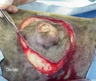
SURGERY
If the tumor can be cured with one or even two surgeries, this is ideal. Mast cell tumors are highly invasive and very deep and extensive margins (at least 3 cm in all directions) are needed. This can be a problem for tumors located on the neck or in the mouth. Further, the inflammation associated with manipulation of the tumor can lead to extra swelling, bleeding, and even a drop in blood pressure. In one study a 10% incidence of wound healing failure ("dehissence") was observed with mast cell tumors. The biopsy sample obtained will not only yield the grade of the tumor but will include a measurement of the tissue margin (the width of normal tissue that has been excised around the tumor). The width of the margins will go far in determining if further treatment is needed. If the margins are narrow or margins indicate there is still tumor left behind then a second surgery or even a course of radiation therapy may be desirable. Clean margins are generally defined as a 10 mm margin around the tumor in all directions. If the margin is clean, theoretically the tumor should be completely removed but it is still a good idea to keep an eye on the area over the years.

CHEMOTHERAPY
Currently three anti-cancer drugs have been particularly helpful in combating mast cell disease: Corticosteroids (such as prednisone), Lomustine, and Vinblastine.
Chemotherapy denotes the administration of certain anti-cancer drugs in order to delay/prevent tumor growth or spread. It may be used before or after surgery, or alone.
Prednisone (a cortisone) is the most commonly used drug for therapy of mast cell tumors. It is well tolerated by dogs and is usually employed for a minimum of six months. If no new tumors appear within that time, your doctor may wean your dog off the prednisone completely. The side effects of prednisone include weight gain, increased appetite and thirst, bladder or skin infections, and panting. Occasionally, stomach irritation or ulcers can occur, or inflammation of the pancreas. Most of the time, the drug dose can be titrated to the patient to minimize any overt symptoms. If the tumor type is determined to be aggressive, additional drugs such as stomach protectants may be prescribed to guard against untoward tumor effects.
Vinblastine Protocol *
For recurrent or multiple tumors, and for those tumors that cannot be surgically removed, combination chemotherapy can be effective in controlling tumor growth and spread for weeks to months or more. A cure per se is generally not realistic, but many dogs tolerate therapy extremely well. The 6-month protocol involves:
- Predinsone: high dose at first, then taper over 4 months
- Vinblastine: an outpatient injection, given once every 21 days
- Cytoxan ®: an oral chemotherapy drug, given by the owners on days 8, 9, 10, and 11 of a 21-day
cycle.
Side Effects
The side effects of prednisone are discussed above. Vinblastine and Cytoxan have the ability to cause nausea and or vomiting, though this is not usual. The most important possible side effects are lowering the bodys defenses so that frequent infections occurs, or (rarely) causing many mast cells to release their contents at once. Both situations can be life threatening. However, these are NOT common, and the risk of these is significantly lower than the risk of untreated mast cell disease. You will be given instructions on what to do if any side effects occur, so do not hesitate to contact us.
The mast cell tumor releases histamine-containing granules that lead to inflammation and increased stomach acid secretion.
These unpleasant symptoms may be alleviated with the use of H1 blocker antihistamines such as Diphenhydramine (Benadryl) and others as well as H2 blocker antihistamines such as Famotidine(Pepcid), Ranitidine (Zantac), Cimetidine (Tagamet) and others.
These medications help palliate the inflammatory effects of the spreading malignant mast cell tumor.